Fig. 1.
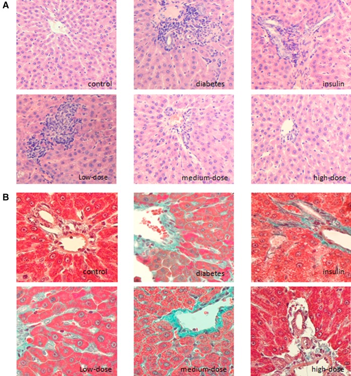
1,25(OH)2D3 exhibited protective effects in livers from diabetic rats. a H&E staining (×200) showed obvious inflammatory cell infiltration around hepatocytes and diffuse infiltration around the portal vein in diabetic rats. Infiltration of inflammatory cells with steatosis of hepatocytes was observed in diabetic rats treated with insulin. Alleviation of the infiltration of inflammatory cells was observed in diabetic rats treated with medium- and high-dose 1,25(OH)2D3. b Masson’s trichrome staining (×400) showed colorization around perisinusoidal spaces, cells, and the portal area, and the structure of the hepatic lobule was abnormal in diabetic rats. The level of interstitial fibrosis decreased, and the structure of the liver was intact in diabetic rats treated with high-dose 1,25(OH)2D3

