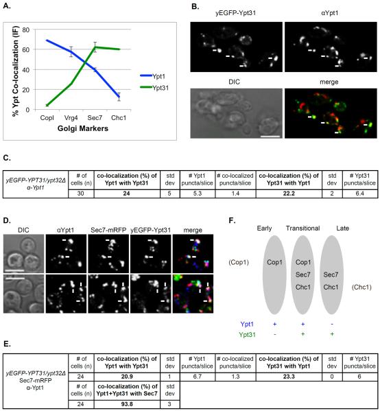
Figure 5. Ypt1 and Ypt31 co-localize on a Sec7-marked Golgi compartment.
A. Both Ypt1 and Ypt31 show intermediate levels of co-localization with Sec7. Summary of IF analyses of Ypt1 (Figure 2B, red line) and Ypt31 (Figure 4B, green line) co-localization with the Golgi markers. B. About 20–25% of Ypt1 and Ypt31 co-localize with each other. Cells deleted for YPT32 and expressing yEGFP-Ypt31 from its endogenous locus were processed for IF microscopy using anti-Ypt1 antibodies (secondary antibody conjugated with red (Texas Red) fluorescent dye). Shown: top panels: Ypt31 (green) and Ypt1 (red); bottom panels: DIC and merge (yellow). C. Table shows quantifications from two independent experiments for panel B; bolded numbers show the co-localization of the two Ypts. D. >90% of the Ypt1-Ypt31 puncta also contain Sec7 in a three-color IF microscopy. Cells deleted for YPT32 and expressing yEGFP-Ypt31 and Sec7-mRFP from their endogenous loci were processed for IF microscopy using anti-Ypt1 antibodies (secondary antibody conjugated with α-Alexa-fluor 647, false colored blue). Shown from left to right: DIC, Sec7, Ypt31, Ypt1 and merge of 3 colors (white). Panels B and D: white arrows point to co-localized signal; bar, 5μm. E. Table shows quantifications from two independent experiments for panel D; bolded numbers show the 2 and 3-color co-localizations. F. Ypt1 and Ypt31 co-localize in a transitional Golgi compartment marked by Cop1, Sec7 and Chc1. A diagram showing three Golgi compartments: early, transitional and late and the distribution of Ypt1 (blue) and Ypt31 (green) in these compartments.

