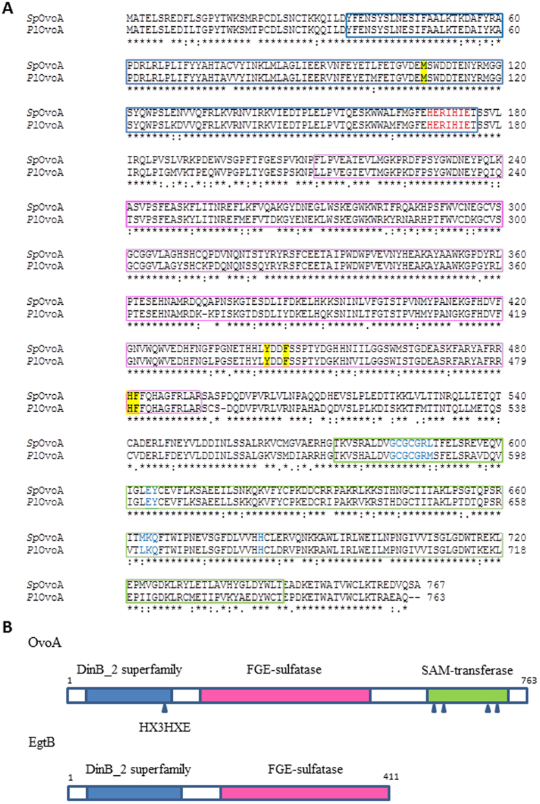
Figure 3. Characterization of OvoA protein in sea urchins.
(A) Sequence alignment of PlOvoA and SpOvoA. DinB superfamily domain (36–176 aa, PlOvoA) in the N-terminal region is boxed in blue. The putative iron binding motif (HX3HXE) is indicated in red. The FGE-sulfatase domain (211–491 aa) is boxed in magenta and the SAM-transferase domain (572–743) in the C-terminal region is boxed in green. The residues (581–587, 602–603, 661–663, 680) belonging to the SAM-binding site are indicated in blue. The putative residues accounting for binding to cysteine and histidine are highlighted in yellow. (B) Schematic representation of OvoA and EgtB primary structure. DinB_2 superfamily domain is boxed in blue and the putative iron-binding site is indicated by an arrow, FGE-sulfatase domain is boxed in magenta. SAM-transferase domain is boxed in green. SAM binding sites are highlighted by arrows.

