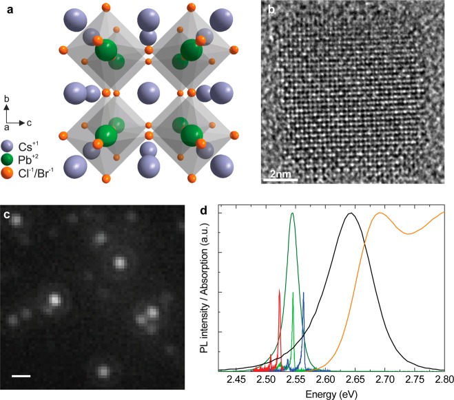
Figure 1.
(a) Illustration of the crystal structure. (b) High-resolution transmission electron microscopy (HR-TEM) image of a single nanocrystal. (c) PL microscopy image from a low-density nanocrystal film obtained with (nonhomogeneous) wide-field excitation. The scale bar corresponds to 2 μm. (d) Absorption (orange) and PL (black) spectra from a high-density ensemble at room temperature, and the corresponding PL (dark green) at T = 6 K. Representative spectra of three individual quantum dots in the low-density film (blue, green, red) at T = 6 K are superimposed.

