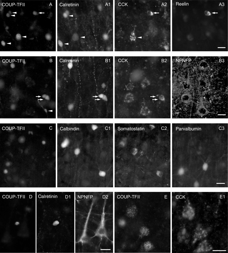
Figure 3.
Immunofluorescence characterization of COUP-TFII expressing using up to 5 antibodies and 4 filter sets on the same cells. (A) Most calretinin-positive neurons (e.g., arrowhead) are COUP-TFII-positive and some of them express CCK (arrowhead) and/or reelin (arrow) in upper layer II. (B) In middle layer III, most calretinin-positive neurons are also COUP-TFII-positive (arrows) and some express CCK (lower arrow). Many pyramidal cells also express CCK in patches representing the Golgi apparatus (B2) and these cells are also immunopositive for NPNFP (B3), but immunonegative for COUP-TFII and calretinin. The small nuclei of some cells in the wall of blood vessels (B, arrowhead) are immunopositive only for COUP-TFII. (C) Interneurons positive for calbindin, somatostatin, or parvalbumin were always immunonegative for COUP-TFII in layers II and III. (D) A calretinin-positive interneuron is also positive for COUP-TFII, but nearby NPNFP-positive pyramidal cells are immunonegative for COUP-TFII in lower layer III. (E) Weakly COUP-TFII-immunopositive large nuclei belong to CCK-positive pyramidal cells in layer VI. The CCK-positive perinuclear patches represent the Golgi apparatus. Scale bar: A–E, 20 µm.

