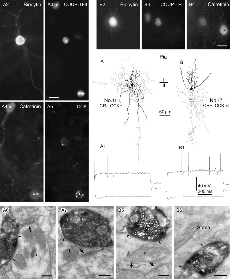
Figure 5.
Interneurons immunopositive for COUP-TFII show irregular firing patterns in vitro and innervate mostly small dendrites. (A and B) Axonal (gray) and dendritic (black) distribution of 2 COUP-TFII-immunopositive cells in layer II. (A1 and B1) Both cells responded with a robust sag to hyperpolarizing current steps (bottom traces), and fired irregular spikes to depolarizing current steps (top traces). (A2–A5 and B2–B4) Cell A was immunopositive for CCK, but negative for calretinin, whereas cell B was positive for calretinin; it was not tested for CCK. Note 2 COUP-TFII-/calretinin-positive interneurons in A3 and 4 (asterisks), one of which was also positive for CCK (double asterisks). In B3 and 4, a rare calretinin-positive interneuron is seen that was immunonegative for COUP-TFII (asterisk). (A6,7 and B5,6) Electron micrographs showing large, type II synaptic junctions (between bars) established by the cells shown in (A) and (B), respectively, with small dendritic shafts (d) and an interneuron soma. The innervated dendrites also receive type I synapses (arrows) from other boutons. Scale bars: A and B, 10 µm; A6,7 and B5,6, 200 nm.

