Figure 1.
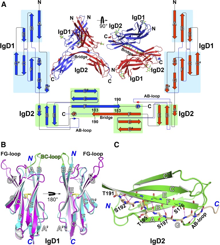
Structure of the PECAM-1 homophilic-binding domain. (A) Side and top views of the structure of IgD1-D2 in the asymmetric unit. N-linked glycans are shown as sticks with green carbons and red oxygens. The backbone hydrogen bonds at the hinge region are dashed in green. The protein topology diagram was generated with Pro-origami43 with manual modifications. The lengths of the β-strand symbols, but not their linkers, are proportional to the lengths of amino acid sequences. Two IgD1-D2 molecules in blue and red form a dimer through swapping of the G β strand at the C terminus of IgD2 domain. (B) Superimposition of PECAM-1 IgD1 (in cyan) and human ICAM-1 IgD2 (in magenta, PDB code, 1IC1), with disulfide bonds represented by yellow sticks. (C) Structure of PECAM-1 IgD2. The swapped G strand is shown in wheat carbons. Oxygens and nitrogens are red and blue, respectively. The hydrogen bonds are dashed in magenta.

