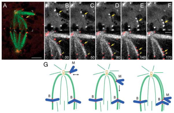
Fig. 2.
Monooriented chromosomes are transported toward the spindle equator along kinetochore fibers of other chromosomes. (A) Two-color fluorescence image of a live PtK1 cell in which kinetochores were labeled with CENP-F/Alexa488 (red) and microtubules with tubulin/rhodamine (green). Area marked with white brackets is enlarged in (B to F). (B to F) selected frames from the two-color time-lapse recording. In each frame, CENP-F/Alexa488 fluorescence (kinetochores) is shown alone (top) and overlaid in red on microtubules (bottom). Arrows mark the kinetochore that moved toward the spindle equator. Note that trajectory of this kinetochore coincided with a prominent kinetochore fiber that extended from the spindle pole to a kinetochore on a bioriented chromosomes already positioned on the metaphase plate (arrowhead). Time in seconds. Scale bars: (A) 5 μm, (F) 2.5 μm. (G) Schematic illustrating the sequence of events presented in (B to F).

