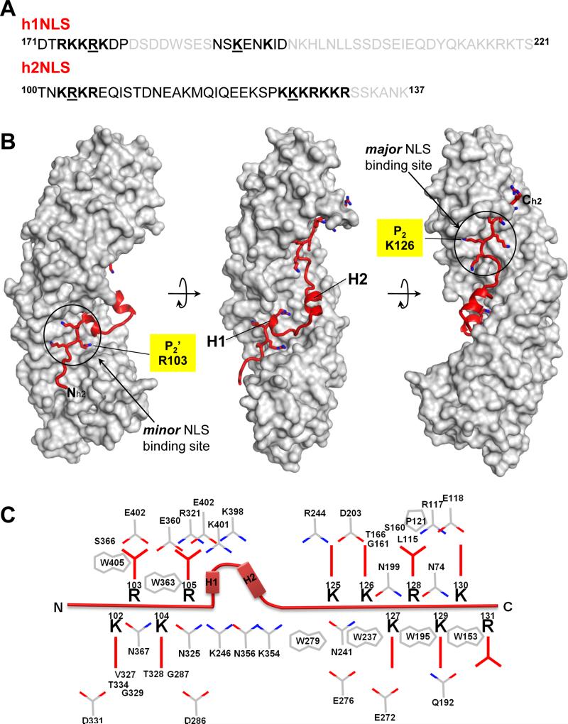
FIGURE 1. Crystal structure of h2NLS bound to ΔIBB-Kap60.
(A) Aminoacid sequence of Heh2 and Heh1 peptides co-crystallized with ΔIBB-Kap60. In black and gray are residues visible and invisible in the crystal structures, respectively; bolded are basic residues occupying minor (left) and major (right) NLS-binding boxes; underlined are residues at position P2’ and P2. (B) Crystal structure of ΔIBB-Kap60 (gray surface) in complex with h2NLS (red ribbon). (C) Schematic diagram of the interactions between h2NLS (in red) and Kap60 residues (in gray) in a distance range of 2.5-4.5 Å. See also Figure S1.

