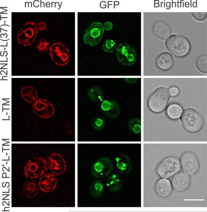
FIGURE 7. In vivo analysis of h2NLS interaction with Kap60.
Deconvolved wide-field images of cells co-expressing Kap60-GFP with mCherry-tagged reporter proteins mCh-h2NLS-L(37)-TM, mCh-h2NLS P2’-L-TM or mCh-L-TM. Scale bar is 5 μm and SEM is indicated. See also Figure S5 and Supplemental Experimental Procedures.

