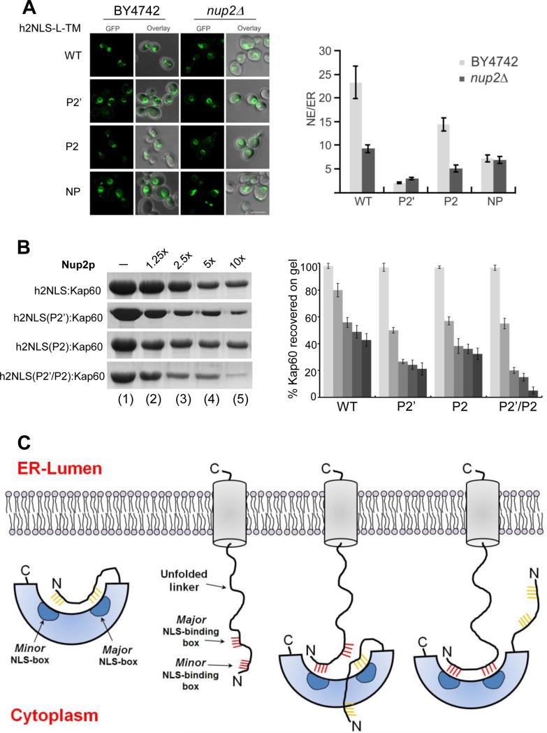
FIGURE 8. Role of Nup2 in displacement of Heh2 from Kap60.
(A) Confocal fluorescence images of a wild type yeast strain (BY4742) and a nup2Δ knockout strain expressing GFP-h2NLS-L-TM and the reporter with mutations at position P2′, P2, and NP-NLS, as well as quantification of average NE/ER-ratios (average of ~30 cells). SEM, and 5 μm scale bar are indicated. (B) Nup2-mediated displacement of ΔIBB-Kap60 from GST-h2NLS (and its mutants at P2’, P2 and P2’/P2) coupled to glutathione beads. The complex was challenged with 1.25-10 fold molar excess of MBPNup2 (res. 1-51) and ΔIBB-Kap60 left on beads is quantified on the right panel (error bars from averaging three independent experiments). (C) Model for recognition and association of a membrane protein NLS to auto-inhibited FL-Kap60. From left to right are schematic illustrations of auto-inhibited FL-Kap60, an ER-synthesized membrane protein (like Heh2) projecting an h2NLS-like import sequence in the cytoplasm and two putative snapshots of FL-Kap60 partially and fully bound to the membrane protein NLS.

