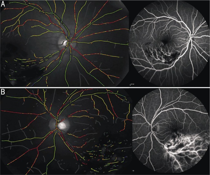
Figure 1. The classification of BRVO based on FFA and oximetry image.

A: The left pseudocolor fundus image of non-ischemic BRVO patient, shows normal retinal oxygen saturation: SaO2-A, 92.8%, SaO2-V, 56.6%; the right FFA image shows the hemorrhage, and non-perfusion area of retinal capillary ≤5 DD; B: The left pseudocolor fundus image of ischemic BRVO patient, shows increased retinal oxygen saturation: SaO2-A, 99.2% SaO2-V, 67.1%; the right FFA image shows the hemorrhage, non-perfusion area of retinal capillary >5 DD, and retinal neovascularization.
