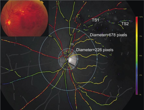
Figure 2. Oxygen saturation map and fundus image of a patient with BRVO.

We performed two kinds of analysis. First kind, the vessel segments between the two circles (approximately central retina) were selected for analysis; Second kind, the first- and second- degree vessels in each four quadrant were selected for analysis. Occluded vessels were strictly chosen.
