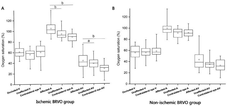
Figure 3. Comparison of oxygen saturation for occluded vessels with unaffected vessels and contralateral eye in BRVO groups.
A: Significantly increased SaO2-A and SaO2-AV can be seen in occluded vessels in ischemic BRVO group (P<0.01), SaO2-V in occluded vessels a little higher than unaffected vessels in ischemic BRVO eye (though no significant difference); B: No significant differences were found between SaO2-V, SaO2-A and SaO2-AV within non-ischemic BRVO group. aStatistically significant difference P<0.05; bStatistically significant difference P<0.01.

