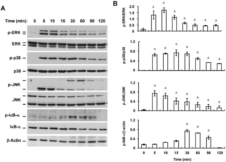
Figure 5. Time-dependent effects of zymosan on MAPK phosphorylation and IκB-α phosphorylation and degradation in HCFs.
A: Serum-deprived cells were incubated with zymosan (600 µg/mL) for the indicated times, after which cell lysates were prepared and subjected to immunoblot analysis with antibodies to phosphorylated (p-) or total forms of ERK1/2, p38, JNK, or IκB-α. β-Actin was examined as a loading control; B: Immunoblots similar to those in (A) were subjected to densitometric analysis to determine the intensity of the bands for phosphorylated forms of ERK1/2, p38, JNK, or IκB-α relative to that of those for the corresponding total forms or β-actin. Data are means±SD from three independent experiments. aP<0.05 (Dunnett's test) versus the corresponding value for time zero.

