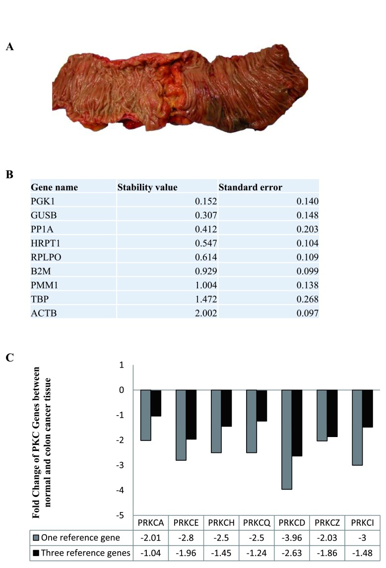
Figure 3. Reference genes in normal and colon cancer tissue.
The stability of the nine candidate reference genes between normal and cancer tissue was analysed using NormFinder. ( A) Surgical image of specimen resected from a colon cancer patient. ( B) Table displaying the stability levels of the nine candidate reference genes between the normal and cancer tissue. ( C) Graph representing the fold change of PKC coding genes in cancer tissue compared to normal tissue (n=21) when using one reference gene (PGK1) versus three reference genes (PGK1, GUSB and PP1A).

