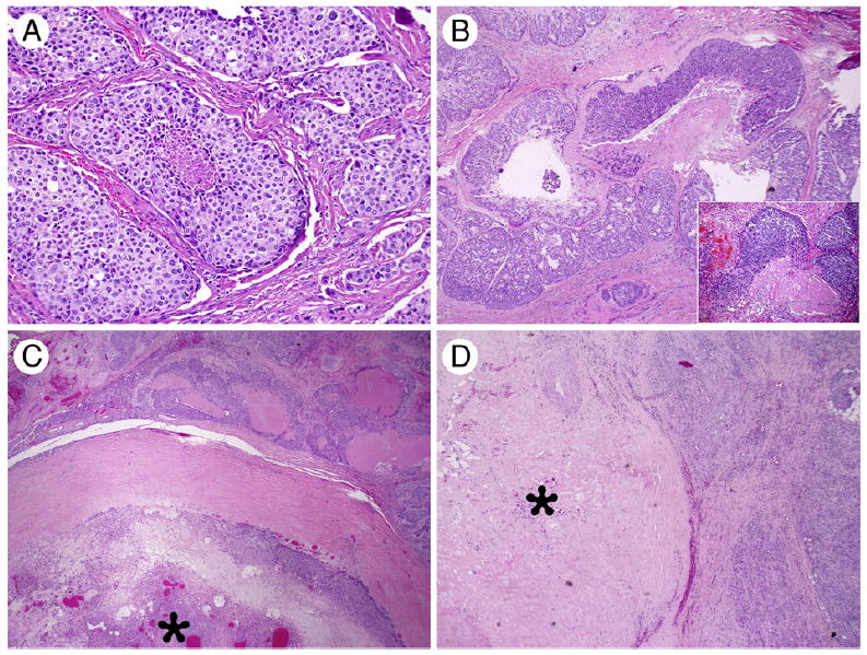
Fig. 1.

Morphologic spectrum of CA ex-PA. A, High-grade SDC type: the tumor cells show abundant eosinophilic cytoplasm and prominent nuclei, arranging in a solid growth pattern with central necrosis (×200). B, MECA with multinodular growth pattern and tumor necrosis (×40) with an inset (×400) showing high-power view of the tumor with hyalinized necrotic stroma and pseudoglandular spaces. C, CA ex-PA associated with a well-demarcated PA component (star; ×20). D, CA ex-PA in which only a discrete hyalinized stromal nodule (star) is noted, most likely representing a burned-out PA (×20).
