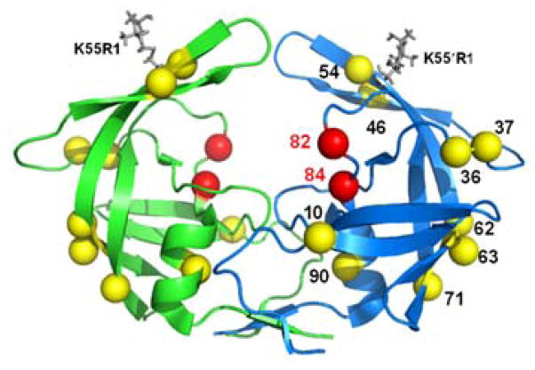
Figure 1.
Ribbon diagram of MDR769 (PDB file 1TW7) protease colored by subunit, rendered in PyMol 1.3. Primary mutations relative to wild-type (LAI) are shown as red spheres, while compensatory mutation sites are rendered as yellow spheres. MTSL spin probes (K55R1) are incorporated in silico via MMM 2011.1 (16) and shown as gray capped sticks.

