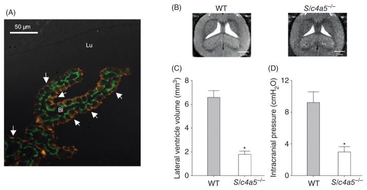
Figure 8.
(A) Double-labeling immunofluorescence microscopic analysis of NBCe2 (red) and NCBE/NBCn2 (green) localization in rat choroid plexus (46). The fluorescence image was overlaid a differential interference contrast image and shows apical localization of NBCe2 (arrows) and basolateral localization of NCBE/NBCn2. Panels B and C show ventricular volume and (D) intracranial pressure in wild-type (WT) and NBCe2 knockout (Slc4a5−/−) mice (152). Panel B shows MRI imaging (horizontal plane) of the lateral ventricles in WT and Slc4a5−/− mice. The ventricular volume and intracranial pressure are significantly reduced in Slc4a5−/− mice.

