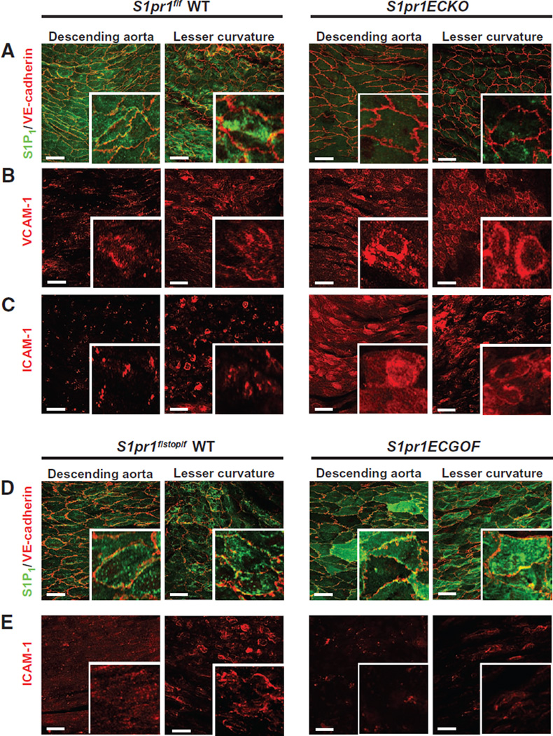
Fig. 2. Genetic modulation of endothelial S1P1 expression and inflammatory marker expression.
Aortae from wild-type (WT) (S1pr1f/f) and S1pr1ECKO (S1pr1f/f Cdh5-CreERT2+/−) mice were dissected, and intima was stained in en face preparations. (A) Immunostaining of S1P1 and VE-cadherin. S1P1 immunofluorescence was undetectable in S1pr1ECKO aortae. (B and C) Immunostaining for VCAM-1 (B) and ICAM-1 (C) in the presence or absence of endothelial S1P1. Images are representative of 10 mice per genotype obtained in 8 to 10 independent experiments. Aortae from WT (S1pr1f/stop/f) and S1pr1ECGOF (S1pr1f/stop/f Cdh5-CreERT2+/−) mice were dissected, and intima was stained in en face preparations. (D) Immunostaining of S1P1 and VE-cadherin. S1P1 immunofluorescence was increased in S1pr1ECGOF aortae. (E) Immunostaining for ICAM-1 with and without endothelial S1P1 overexpression. Images are representative of 10 mice per genotype obtained in five to seven independent experiments.

