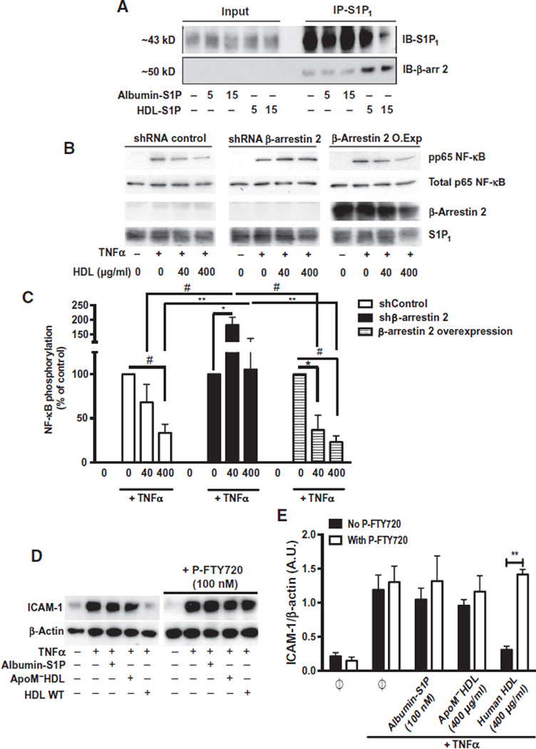
Fig. 6. The role of β-arrestin 2 in the biased signaling of HDL-S1P.
(A) Serum-starved HUVECs were treated with albumin-S1P or human HDL-S1P for the indicated times. Cell lysates were subjected to immunoprecipitation for S1P1 and immunoblotted (IB) with β-arrestin 2 antibody. Immunoblots are representative of three independent experiments. (B) HUVECs stably overexpressing β-arrestin 2 complementary DNA or an shRNA directed against β-arrestin 2 were serum-starved and stimulated with TNFα, with or without human HDL at the indicated doses. p65 phosphorylation was analyzed by immunoblot assay. (C) Data represent combined analysis of p65 phosphorylation from four independent experiments. *P < 0.05, **P < 0.01, #P < 0.005 by two-way ANOVA followed by Dunnett’s multiple comparison. (D) Serum-starved HUVECs were stimulated with TNFα in the absence or presence of albumin-S1P or ApoM−HDL or human HDL as indicated. In the P-FTY720–treated experiments, serum-starved HUVECs were pretreated with P-FTY720 in the absence or presence of albumin-S1P, ApoM−HDL, or human HDL before TNFα stimulation in the presence of P-FTY720. ICAM-1 abundance was assessed by immunoblotting. (E) Data represent the combined analysis of ICAM-1 abundance from three independent experiments. **P < 0.01 by unpaired Student’s t test.

