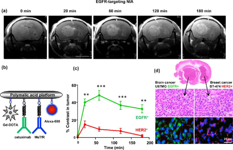
Figure 4.

Detection of EGFR+ GBM in the presence of HER2+ metastatic tumor (double tumor model) using EGFR-targeting NIA. (A) MRI brain scans of mice with double tumors, a GBM (U87MG, EGFR+) in the left hemisphere and metastatic breast cancer (BT-474, HER2+) in the right hemisphere after iv injection of targeted NIA (P/Gd-DOTA/cetuximab/MsTfR-mAb/Alexa-680). (b) Schematic presentation of dual modality (MRI contrast and optical) imaging agent. Gd-DOTA is attached via amide linkage. Maleimide-functionalized mAbs (cetuximab and MsTfR) and optical agent (Alexa-680) are attached by a stable thioether bond. Cetuximab is used for active tumor targeting, and MsTfR mAb is used for endothelial targeting and transcytosis. (c) Quantitative analysis of MRI contrast in tumors. High contrast in the targeted GBM tumor was maintained beyond 3 h, whereas contrast in the nontargeted HER2+ tumor was low and approached the baseline after 2–3 h (**p < 0.01; ***p < 0.001). (d) Confirmation of the MRI diagnosis by immunohistochemical analysis. Brain sections indicated the presence of two tumors by H&E staining. Brain areas identified by MRI as GBM stained positively with anti-EGFR (green) antibodies, and areas not identified by MRI as EGFR+ stained with anti-HER2 (red) antibodies.
