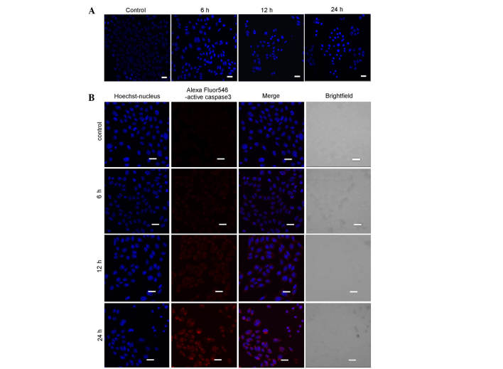
Figure 2.
Myricetin induces apoptosis in SKOV3 cells. (A) The cells were treated with 40 µg/ml myricetin for 0, 6, 12 and 24 h, and were subsequently stained with Hoechst 33342. Confocal microscopy was used to observe cell morphology (scale bar, 20 mm). (B) The cells were treated with 40 µg/ml myricetin for 0, 6, 12 and 24 h. Nuclear staining and fluorescence of active Caspase 3 was observed using confocal microscopy (scale bar, 20 µm).

