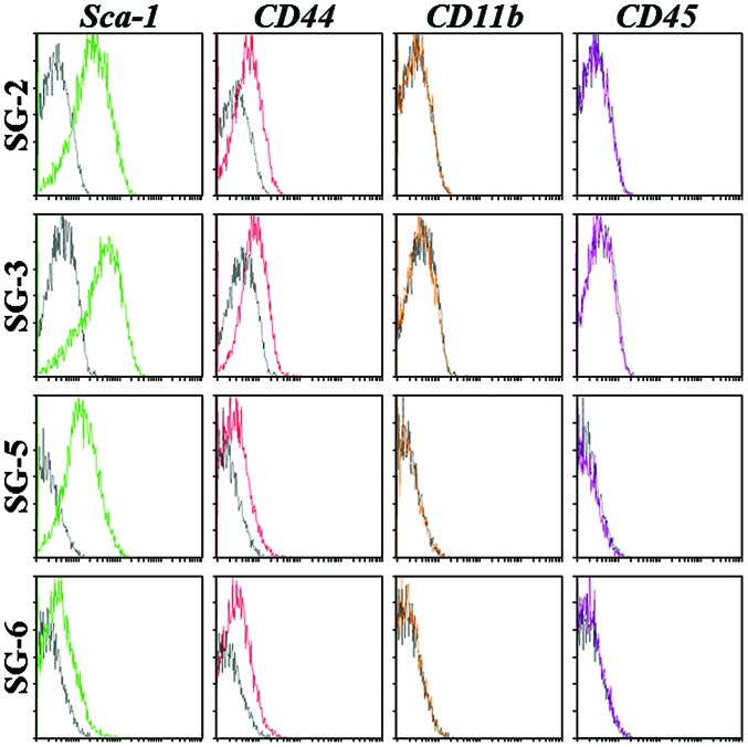
Figure 3.
Identification of mouse mesenchymal stem cell markers in SG-2, -3, -5, and -6 cell by flow cytometry. The cell lines were incubated with phycoerythrin-conjugated control immunoglobulin G (black), anti-Sca-1 (green), anti-CD44 (red), anti-CD11b (yellow), or anti-CD45 (purple) antibody and acquisition was performed on a EPICS XL ADC system.

