
Figure 1.
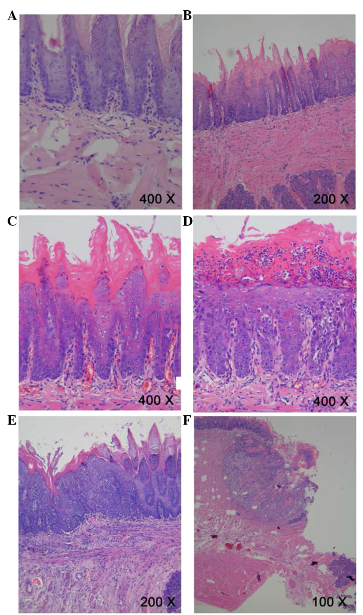
Pathological evidence of carcinogenesis in rat tongues. (A) Normal squamous epithelium of the tongue. Magnification, ×400. (B) Mild epithelial dysplasia of the tongue exhibited loss of polarity of the deeper cell layers of the epithelium and mild nuclear pleomorphism of cells. Magnification, ×200. (C) Moderate epithelial dysplasia of the tongue indicated a basaloid appearance, loss of polarity of cells and intercellular cohesion. Magnification, ×400. (D) Severe epithelial dysplasia of the tongue exhibited a proliferation of basal cells, grossly disturbed stratification, the loss of polarity of cells, individual cell keratinization and nuclear pleomorphism of cells. Magnification, ×400. (E) Carcinoma in situ of the tongue exhibited the proliferation of primitive basal epithelial cells from the basement membrane to the surface, marked nuclear atypia and the full thickness of the epithelium. Magnification, ×200. (F) Squamous cell carcinoma of the tongue showed gross disruption of normal epithelial architecture, prominent cellular pleomorphism, the formation of dyskeratosis with keratin pearl and invasion into underlying connective tissues. Magnification, ×100.