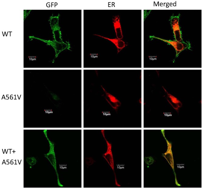
Figure 5.
Representative images of subcellular localization of the hERG protein expressed in HEK-293 cells. Top panel, HEK-293 cells expressing pEGFP-C2-WT; middle panel, HEK-293 cells expressing pEGFP-C2-A561V; and bottom panel, HEK-293 cells expressing pEGFP-C2-WT and pEGFP-C2-A561V. Left column, the hERG protein tagged with green fluorescence (green); middle column, HEK-293 cells transfected with pDsRed2-ER (red); and right column, merge of the two. Scale bar, 10 µm. GFP, green fluorescent protein; ER, endoplasmic reticulum, WT, wild-type.

