Figure 1.
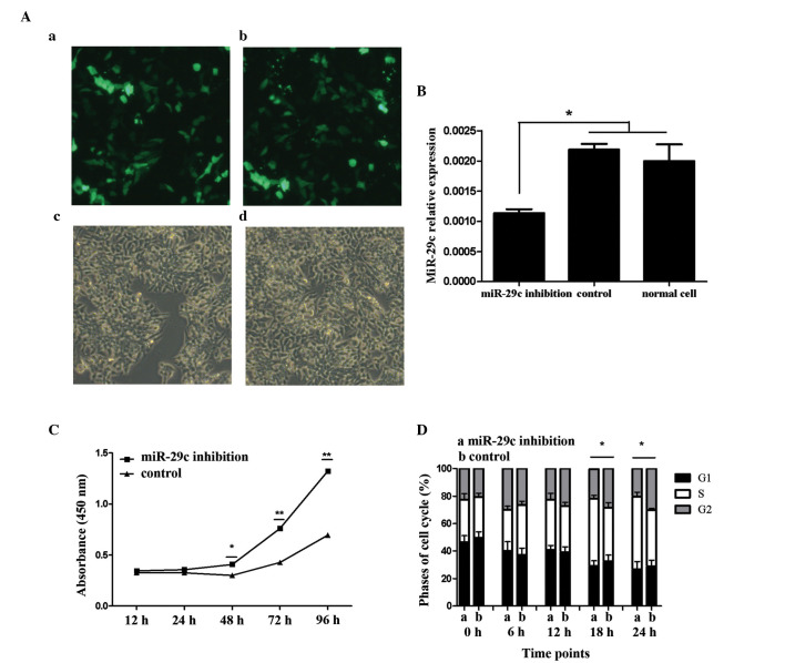
Expression of miR-29c in P19 cells and promotion of P19 cell proliferation by miR-29c. Transfection efficiency was determined by the expression of green fluorescent protein using fluorescence microscopy. (A) (a) miR-29c inhibition and (b) control plasmids were successfully transfected and expressed. No significant differences in transfection efficiency were found between the (c) miR-29c-inhibited and (d) control groups (magnification, x100). (B) Confirmation of the expression of miR-29c by reverse transcription-quantitative polymerase chain reaction analysis. Data are presented as the mean ± standard deviation of three experiments. miR-29c was significantly downregulated in the miR-29c inhibition group, compared with the control- and no-vector P19 cell group (*P<0.05). Expression levels of miR-29c were similar between the control- and no-vector P19 cell groups, indicating the vector itself exerted no effect. (C) Cell proliferation was monitored over 4 days consecutively using a Cell Counting Kit-8 assay, performed three times with six replicates in each group. Representative proliferation curves are shown (*P<0.05 and **P<0.01). (D) Cell cycle analysis of the P19 cells was performed using flow cytometry to detect proliferation. Data are presented as the mean ± standard deviation of three experiments (*P<0.05). Results from the two assays indicated that miR-29c inhibition promoted P19 cell proliferation. miR, microRNA.

