Figure 1.
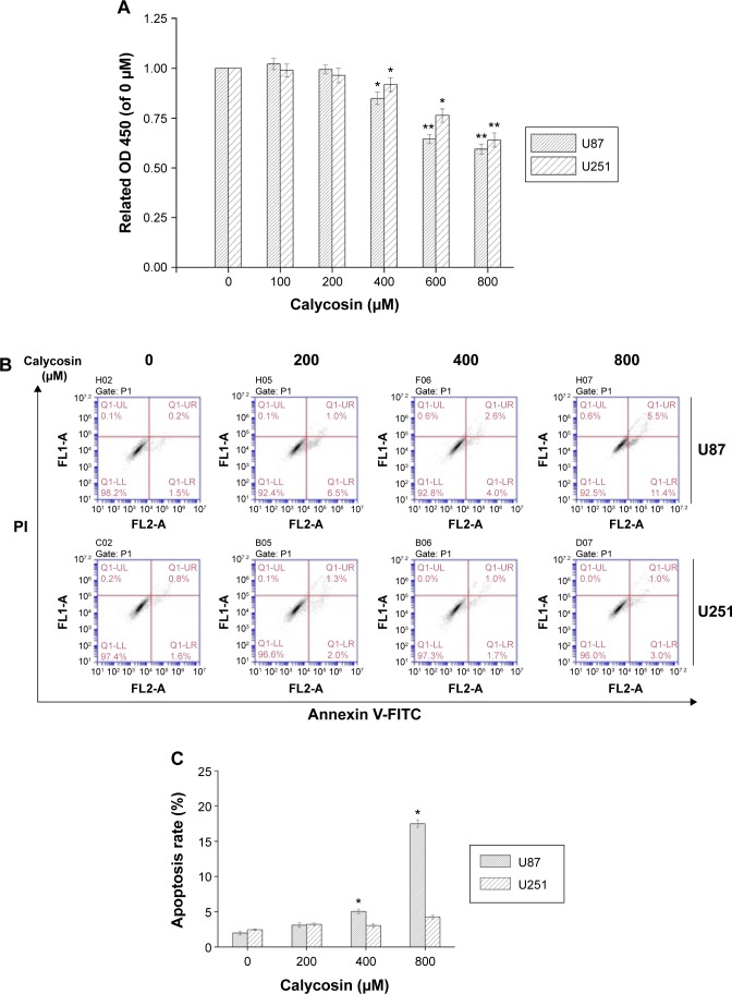
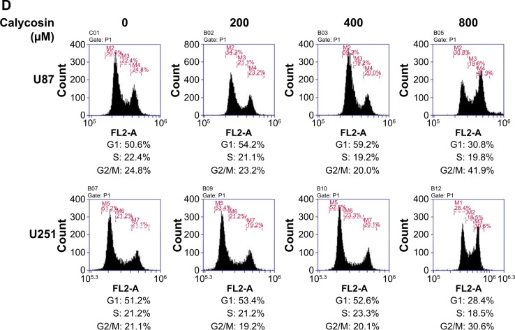
Effects of calycosin on proliferation, apoptosis, and cell cycle regulation in U87 and U251 cells.
Notes: (A) Cells were treated with various concentrations of calycosin for 24 hours before MTT assay. (B and C) Cells were treated with the indicated concentrations of calycosin (0, 200, 400, and 800 μM) for 24 hours and stained with Annexin V-FITC/PI. At least 10,000 cells were tested per sample. All tests were performed in triplicate and presented as mean ± standard deviation. *P<0.05, **P<0.01, compared with control (0 μM). (D) Cells were exposed to different concentrations of calycosin for 24 hours. After treatment, cells were harvested and subjected to flow cytometric analysis to assess the cell cycle distribution. At least 10,000 cells were analyzed per sample. After treatment, cells were harvested and subjected to flow cytometric analysis to assess the cell cycle distribution. At least 10,000 cells were analyzed per sample.
Abbreviations: OD, optical density; PI, propidium iodide.


