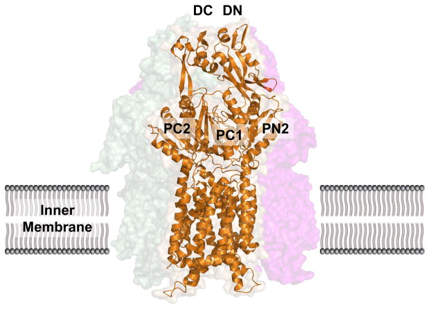
Fig. 3. Crystal structure of the apo-CusA efflux pump.
The CusA protomer found in the asymmetric unit of the crystal lattice is depicted by ribbon diagram (orange). The surface rendering corresponds to the CusA homotrimer, which is formed by symmetry within the crystal. Sub-domains PN1, PN2, PC1 and PC2 form the pore domain, while subdomains DN and DC comprise the docking domain, presumably interacting with the CusC channel.

