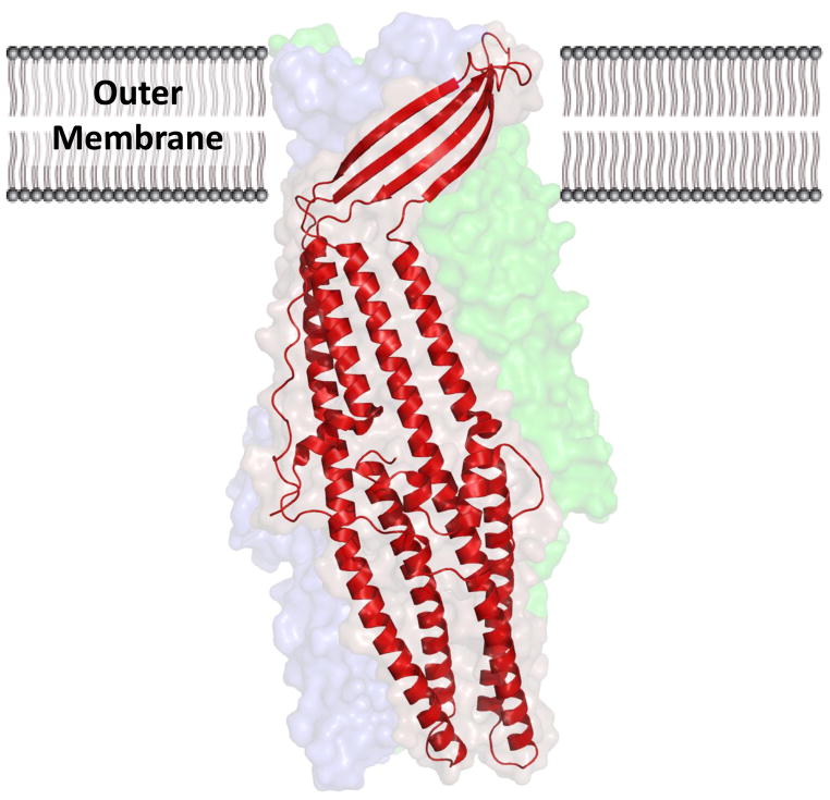
Fig. 9. Crystal structure of the CusC outer-membrane transporter.
The CusC monomer (red) within the asymmetric unit of the crystal is depicted by ribbon diagram. The surface rendering corresponds to the biological CusC trimer, which is created by crystal symmetry. Each subunit of CusC consists of a four β-strands atop nine α-helices, which are arranged as a barrel in the trimeric structure.

