
Figure 5.
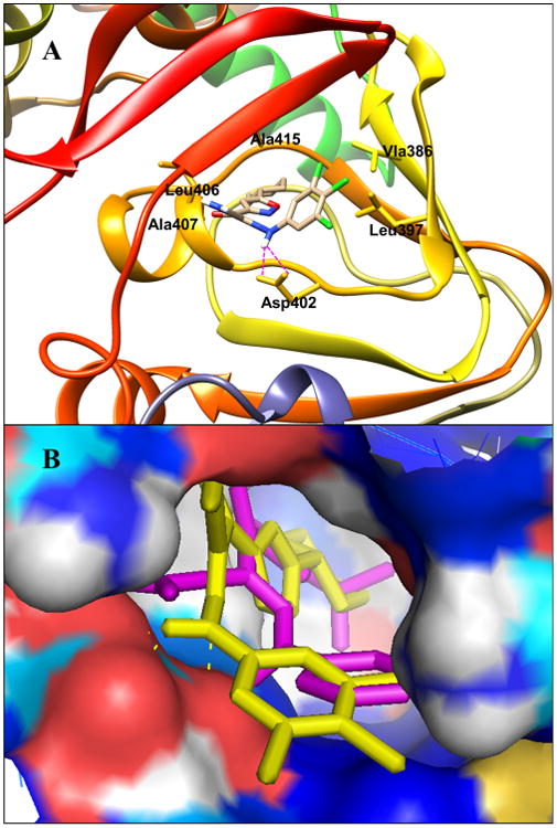
(A) Predicted binding mode and molecular docking of 47 into the cAMP binding domain B (CBD) of EPAC2 protein. Important residues are drawn in sticks. Hydrogen bonds are shown as dashed purple lines. (B) Overlay analysis of ligands 1 and 47. 1 is shown in purple, and 47 is depicted in yellow.