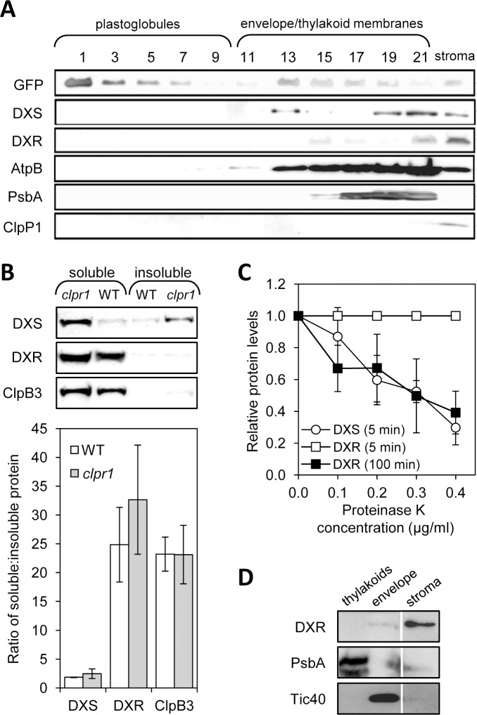
Fig 1. Immunoblot analysis of chloroplast subfractions and protein stability.
(A) Chloroplasts isolated from transgenic plants overexpressing PGL34-YFP were used to separate soluble (stromal) and membrane fractions. The latter were loaded in a sucrose density gradient and separated by ultracentrifugation. Proteins contained in 35 μl of sequential fractions collected from the top of the gradient or from the stromal sample were separated by SDS-PAGE and transferred to a membrane for immunoblot analysis with antibodies against GFP (to detect PGL34-YFP) or the indicated endogenous proteins. (B) Immunoblot analysis of the distribution of DXS and DXR proteins in soluble and insoluble fractions isolated from native protein extracts of wild type and clpr1 mutant seedlings. The graph represents mean ± SEM of the ratios of soluble vs. insoluble protein levels in n = 3 independent experiments. (C) Quantification of DXS and DXR protein levels after immunoblot analysis of wild type protein extracts incubated with the indicated concentrations of proteinase K for the indicated times. Mean ± SEM of n = 4 independent experiments are shown. (D) Immunoblot analysis of the distribution of DXS in envelope and thylakoid membranes isolated from wild type chloroplasts. A lane corresponding to the stromal fraction is also shown. The same extracts were incubated with antibodies against marker proteins of the envelope (Tic40) and the thylakoid membranes (PsbA). Representative blots are shown in all cases.

