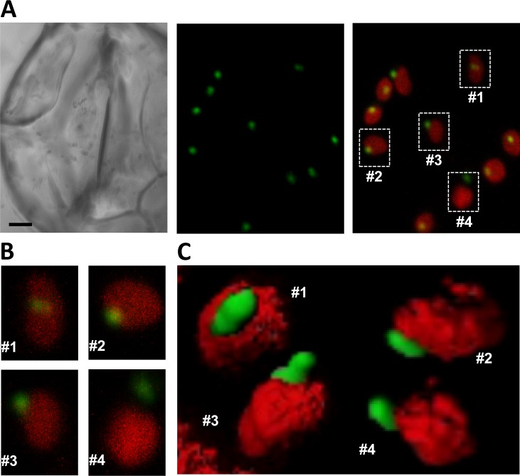
Fig 5. Differential localization of DXR-GFP bodies inside and outside chloroplasts.
(A) Guard cells of a 35S:DXR-GFP (H line) plant. The pictures show a bright field image (left panel), GFP fluorescence (central panel), and merged GFP and chlorophyll fluorescence (right panel). Bar, 2 μm. (B) Magnification of the chloroplasts boxed in (A). (C) Reconstructed 3D images of representative chloroplasts at the indicated phases of DXR-GFP vesicle development.

