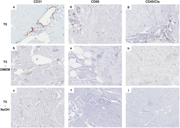
Fig 6. Immunohistochemical reactions showing histological features of the cellular component of reticular dermal matrices at different incubation times (20X magnification).
In control samples (T0), the CD31 immunohistochemical reaction showed the presence of intact vessels (A; endothelial cells are stained in brown), while immunostaining was focal and weak after 5 weeks (T5) of treatment with DMEM (B) and completely absent in NaOH treated samples (C). Immunohistochemistry also showed that at T0 both macrophages (D, showing CD68 positive cells) and lymphocytes (G, showing CD45/CLA positive cells) were present, concentrated in perivascular spaces and near the residual lower portion of hair follicles, while only non-specific focal and weak staining was present after 5 weeks (T5) of treatment with DMEM (E, H); reactions were completely negative in NaOH-treated samples (F, I).

