
Fig 7. Bright field imaging.
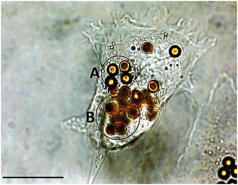
Bright field microscopic image showing a single fibroblast in the middle of the image as the host for several MB. The focus of the image reveals the cell body including its periphery and some of the beads that can be seen in focus (e.g., circle A) than the remaining MB. Other beads are not in the focal plane (e.g., circle B) indicating two different planes of the vertical positions of MB. Scale bar indicates 20 μm.