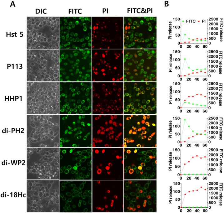
Fig 5. Localizations of FITC-peptides in Candida albicans cells.
C. albicans cells were incubated for 10 minutes with 10 μg/ml of FITC-peptides and visualized by confocal microscopy. The left panel shows DIC fluorescent and FITC-PI merge images of Candida cells with FITC-peptides and PI (5 μg/ml). The right panel shows FITC-peptides and propidium iodide uptake assay. FITC-peptides and PI uptake was measured for 60 min in Perkin Elmer 2030 reader (VictorX 3).

