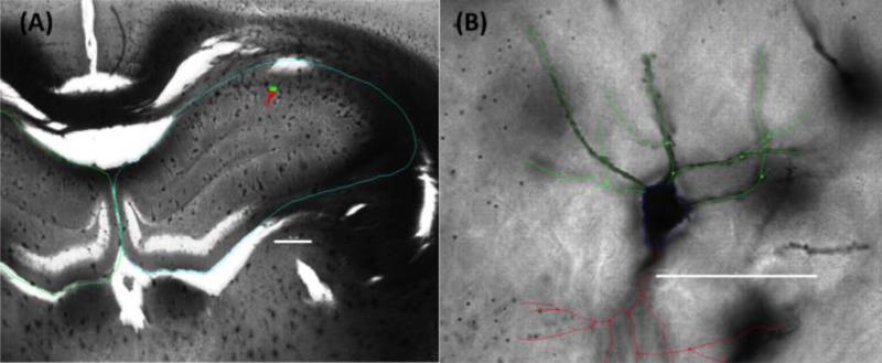
Figure 5.
Representative photomicrographs of a Golgi-Cox stained section of dorsal (A) hippocampus (blue tracing) at low magnification, highlighting the location of a prototypical CA1 pyramidal neuron (green/red marker) and (B) a high magnification view of the same cell with basal (green) branch tracings used for analysis. Apical (red tracing) branches are included for orientation purposes. Scale bars are (A) 500μm and (B) 50μm.

