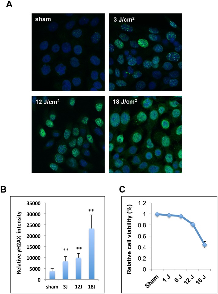
Fig 1. H2AX phosphorylation and cell viability after ssUV irradiation.
(A, B) ssUVR induces γH2AX in human keratinocytes. HaCaT cells were exposed to ssUVR at 0, 3, 12, 18 J/cm2, the levels of γH2AX was determined at 1 hour after irradiation. (A) Representative image. (B) Relative γH2AX intensity in irradiated HaCaT cells. γH2AX intensity was quantified as the corrected total cell fluorescence and presented as the mean ± SD (n = 54). Statistical significance of the difference between sham and ssUVR-treated cells was analyzed using Student's t-test. ** p<0.01 (C) HaCaT cells were exposed to various doses of ssUVR (0, 1, 6, 12, and 18 J/cm2). Cell viability was assessed at 24 hours after exposure, and presented as the percentage of viable cells relative to the total cell numbers. The results showed the mean ± SD (n = 4)

