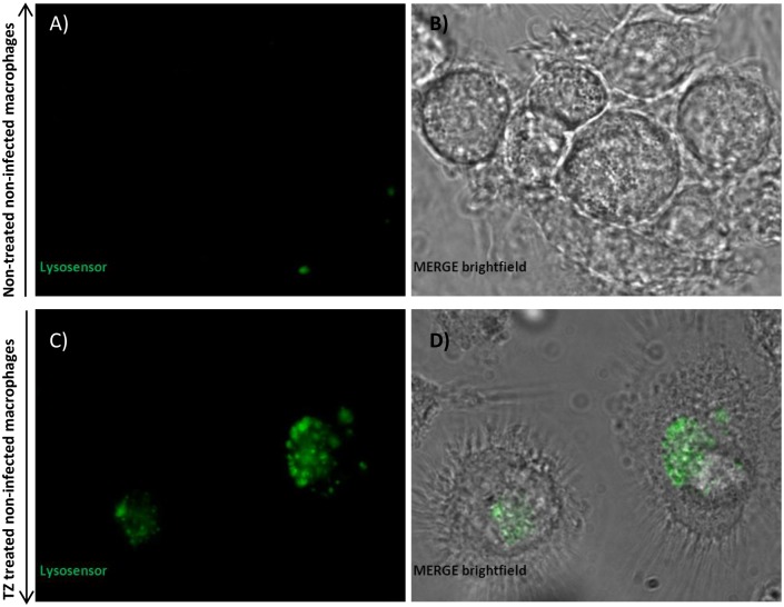
Fig 7. Qualitative analysis of phagosomal acidification in macrophages stained with LysoSensor Yellow/Blue by confocal microscopy.
Evaluation of fluorescence intensity of (A) non-infected non-treated macrophages and (B) brightfield for non-infected non-treated macrophages; evaluation of fluorescence intensity of (C) non-infected macrophages treated with thioridazine (TZ) and (D) brightfield for non-infected macrophages treated with thioridazine, with the aid of the fluorescent probe LysoSensor Yellow/Blue. LysoSensor Yellow/Blue DN160 emits yellow/dark green fluorescence at neutral pH and blue/green fluorescence at pH 5–5.5. Data was analysed using a LSM 510 META laser scanning confocal microscope.

