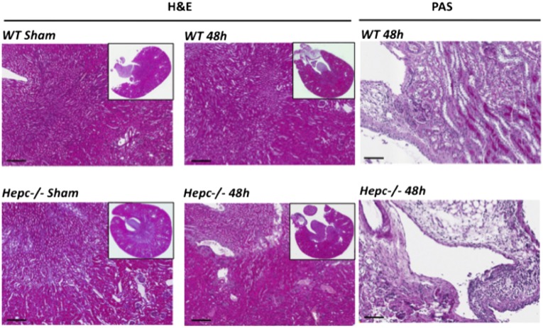
Figure 2.
Histologic analysis in the kidney. WT and Hepc−/− mice were infected with 109 colony-forming units of CFT073. After 48 hours postinfection, kidney sections were stained with hematoxylin and eosin staining (H&E) and with periodic acid–Schiff staining (PAS). (Sham) means uninfected mice. Scalebars represents 200 µm for H&E and 80 µm for PAS.

