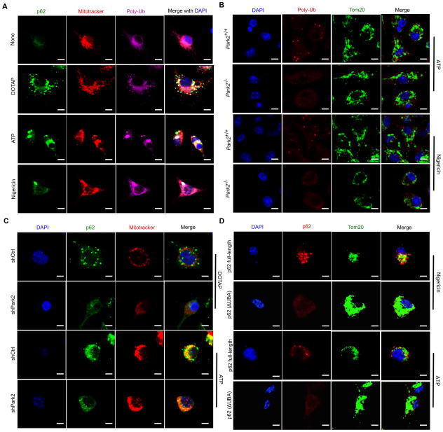
Figure 3. Mitochondrial p62 recruitment is Parkin dependent.
(A) Intracellular distribution of p62, polyubiquitin (poly-Ub) and mitochondria (Mitotracker) in LPS-primed WT BMDM stimulated with NLRP3 agonists determined by confocal microscopy. Scale bars: 5 μm. (B) Mitochondrial poly-Ub decoration examined by confocal microscopy in LPS-primed Park2+/+ or Park2−/− BMDM stimulated with NLRP3 agonists. Scale bars: 5 μm. (C) Mitochondrial recruitment of p62 in LPS-primed WT (shCtrl) or Parkin-deficient (shPark2) iBMDM stimulated with NLRP3 agonists. Scale bars: 5 μm. (D) Subcellular distribution of human p62 in LPS-primed, NLRP3 agonist stimulated p62-deficient BMDMs transduced with human p62-full length or p62(ΔUBA) constructs. Scale bars: 5 μm.

