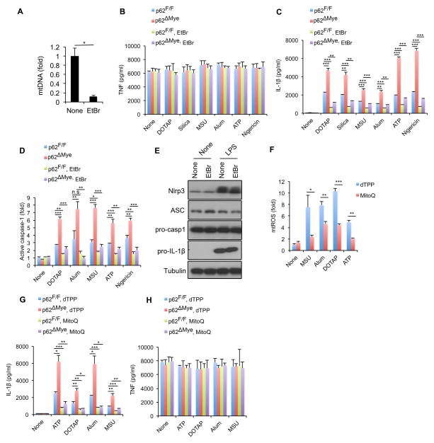
Figure 6. Elimination of mitochondrial signals prevents excessive IL-1β production by p62-deficient macrophages.
(A) Relative concentrations of mtDNA in p62F/F and p62ΔMye BMDM before and after 4 days of ethidium bromide (EtBr) treatment. Results are averages ± s.d. (n=3). (B–D) TNF (B) IL-1β (C) release and caspase-1 activity (D) in p62F/F and p62ΔMye BMDM pre-treated with EtBr or not that were co-stimulated with LPS and different NLRP3 agonists. Results are averages ± s.d. (n=3). (E) IB analysis of pro-IL-1β, NLRP3, ASC, and pro-caspase-1 in lysates of EtBr-pretreated WT BMDM before and after 4 hrs of LPS stimulation. Data are representative of three independent experiments. (F) Relative mtROS amounts determined by MitoSOX staining of LPS-primed WT BMDM stimulated with NLRP3 agonists in the presence of dTPP or Mito-Q. (G,H) Release of IL-1β (F) and TNF (G) from LPS-primed p62F/F and p62ΔMye BMDM; treated with Mito-Q or vehicle (dTPP) before addition of NLRP3 agonists. Results are averages ± s.d. (n=3).

