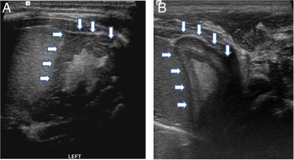
Figure 2.

Ultrasound of the adrenal gland obtained on the 10th day, compared with a normal scan. (A) Transverse scan shows enlargement of the adrenal gland with ‘cerebriform’ appearance, the wrinkled surface resembles the appearance of brain gyri. (B) Normal adrenal gland of a healthy infant at the same age. The gland is relatively large and easy to see, the cortex is thick and hypoechoic, whereas the medulla is relatively thin and hyperechoic.
