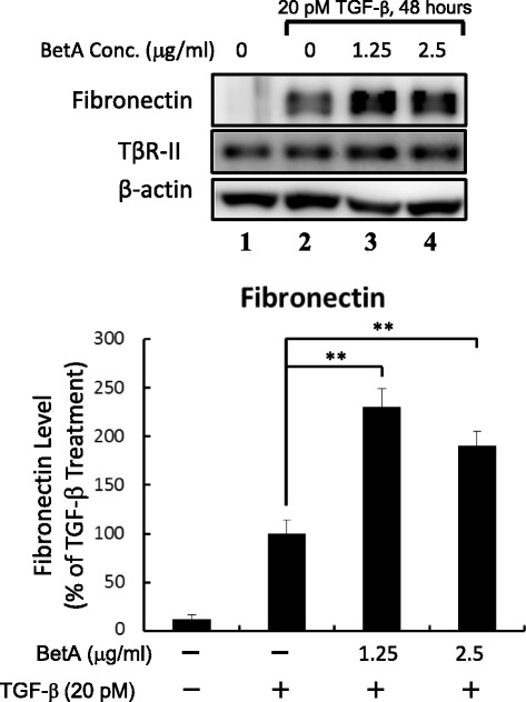
Fig. 6.

BetA promotes TGF-β-induced fibronectin expression. Cells were incubated with 1.25 μg/mL and 2.5 μg/mL of BetA, and then treated with 100 pM TGF-β for 48 h. Whole-cell extracts were prepared and analyzed by Western blot analysis using an antibody against fibronectin (top). The quantitative data from three analyses is shown as mean ± SD (bottom). **The group of BetA/TGF-β co-treatment significantly higher than that in cells treated with TGF-β only (P < 0.01)
