Fig. 2.

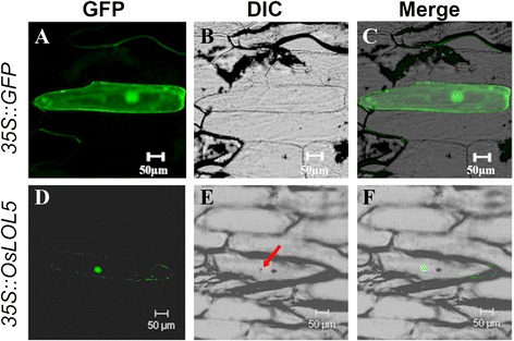
Subcellular localization of the 35S::OsLOL5::GFP fusion protein. a, d GFP, green fluorescence. b, e DIC, bright field. c, f Merge, green fluorescence, and bright field superposition. Nucleus is marked with the red arrow

Subcellular localization of the 35S::OsLOL5::GFP fusion protein. a, d GFP, green fluorescence. b, e DIC, bright field. c, f Merge, green fluorescence, and bright field superposition. Nucleus is marked with the red arrow