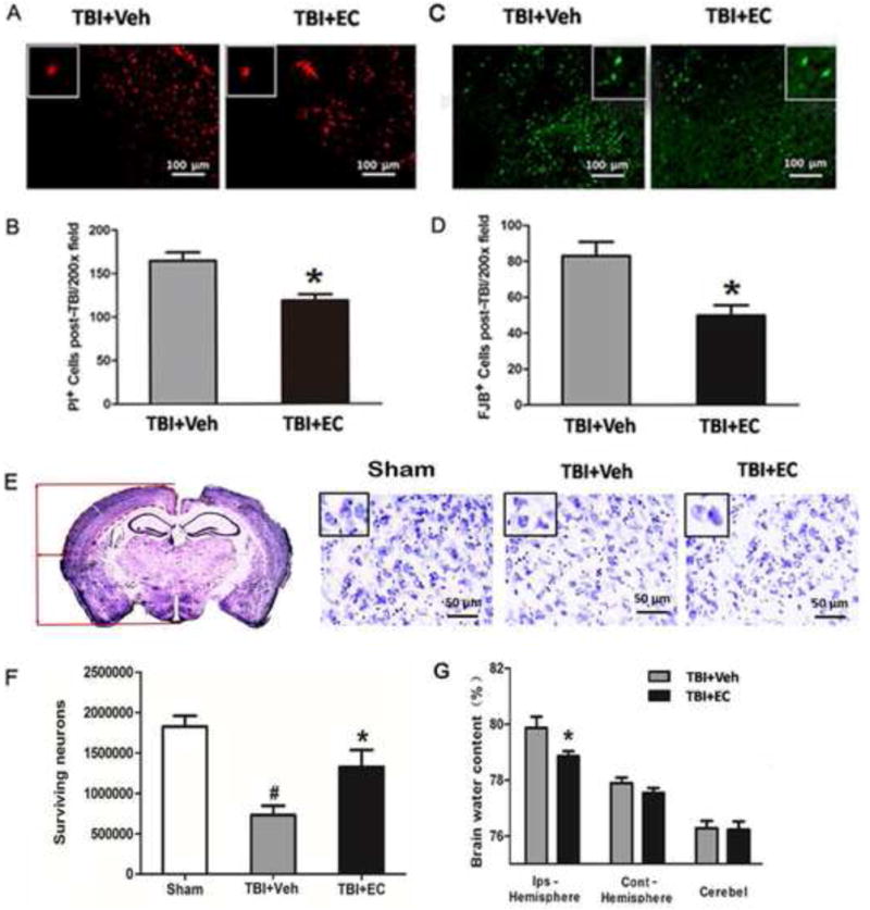
Fig. 3.

EC reduces cell death and neurodegeneration, increases cell survival, and decreases brain water content after TBI. (A) Representative propidium iodide (PI)-stained brain sections are shown for the vehicle-treated and the EC-treated groups. (B) EC decreased the number of PI-positive cells per 200× field. n=6/group, *p<0.05 (t-test). (C) Fluoro-Jade B (FJB)-stained brain sections. (D) EC-treated mice had significantly fewer FJB-positive cells per 200× field than did vehicle-treated mice. n=6/group, *p<0.05 (t-test). (E) Left: a representative Cresyl violet-stained brain section on day 28 after TBI. The location in the cortex used to determine the density of surviving neurons by stereology is delineated by a dashed red line. Right: three representative images of Cresyl violet-stained brain cortical sections from the sham group, the TBI+vehicle group, and the TBI+EC group. Insets represent higher magnification images of Cresyl violet-stained cells in the cortex. Scale bar, 50 μm. (F) Quantitative stereological analysis showed significantly more surviving neurons in the EC-treated mice than in the vehicle-treated mice on day 28 after TBI. n=7/group, *p<0.05 versus the vehicle-treated group; #p<0.05 versus the sham group (one-way ANOVA followed by Newman-Keuls test). (G) At 3 days post-TBI, brain water content of the ipsilateral hemisphere (but not contralateral hemisphere or cerebellum [cerebel]) was significantly lower in the EC-treated group than in the vehicle-treated group. n=8/group, *p<0.05 (t-test).
