
Fig. 1.
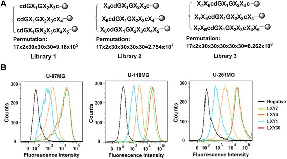
Structures of three focused OBOC libraries and binding affinity of various peptide ligands for α3β1 integrin-expressing glioblastoma cells by flow cytometry. The focused OBOC libraries were designed and synthesized (a). Three glioblastoma cell lines U-87MG, U-118MG, and U-251MG were incubated with biotinylated LXY1 (blue curve), LXY4 (orange curve), LXY7 (green curve), LXY30 (red curve), or negative control (black curve) and then analyzed with flow cytometry (b)