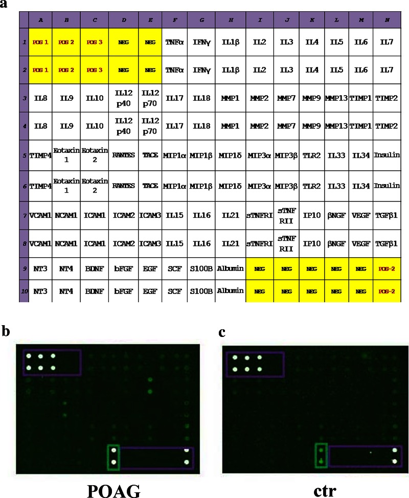
Fig. 2.
Representative chip-based arrays. Chip are grids that contain small amounts of purified proteins in high density, hybridized to sample and detected by fluorescent technique. a The map showing the selected factors in a 14 × 10 grid. Each subarray contains 60 antibodies and specific positive/negative referring spots. b, c Representative GenePix acquired arrays from POAG (b) and post-mortem (c) TM specimens, both loaded as normalized extracts. White points are positive technique controls (framed), dark points are negative technique controls and green points are POAG/post-mortem TMs (cy3-labeled spots). Abbreviations of the main factors were according to International Classification. EGF epidermal growth factor, IL interleukin, MIP macrophage inflammatory protein, MMP matrix metalloproteinase, POAG primary open angle glaucoma, SCF stem cell factor, SDS sodium dodecyl sulphate, TIMP tissue inhibitor of metalloproteinase, TLR toll-like receptor, TM trabecular meshwork, TNF tumor necrosis factor

