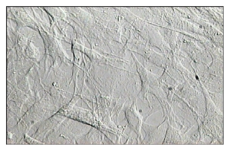
Figure 5.

Mesenchymal Wharton's jelly stem cell. Dissociated mesenchymal Wharton's jelly stem cells were dispersed in 10% FBS-DMEM and counted under a microscope with the aid of a hemocytometer. The mesenchymal cells were then used directly for cultures or stored in liquid nitrogen for later use. 24 hours after injury marked HUMSCs (3 × 10⁵ cells/μL) with BrdU in 9 μL of normal saline were sucked in to a Hamilton syringe and were injected slowly at a rate of 0.25 μL/min by microinjector to 3 separate places and transplanted into the three sites of lesion area (epicenter, distal, and proximal) at a depth of 1.2 mm.
