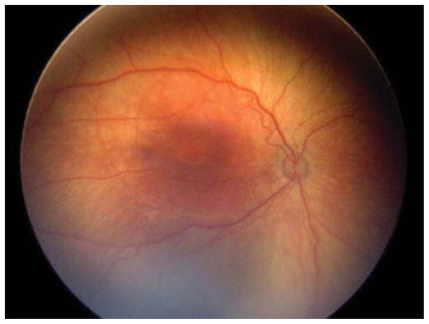
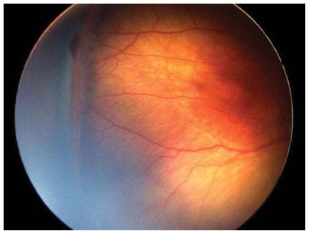
Figure 4. Representative color fundus photograph of a patient (Case 7) treated for retinopathy of prematurity milder than Type 1 disease due to concerning structural changes including tangential traction and temporal vessel straightening and thick stage 3 membrane with anteroposterior traction concerning for progression to stage 4 retinopathy of prematurity, as well stage 3 retinopathy of prematurity too active for his advanced postmenstrual age.
Fundus photos of the right eye (left: posterior, right: temporal) reveal zone II, stage 3 retinopathy of prematurity without evidence of plus disease. There was temporal vessel straightening as well as thick fibrovascular stage 3 with anteroposterior traction.


